Atlas · Plate II · Scientific Basis

Dental Anatomy and Tooth Morphology

Tooth morphology is the shape of every permanent and primary tooth: crown outline, cusps, grooves and, above all, roots. The area also covers numbering systems, eruption timing, occlusion and developmental anomalies. For a hygienist the roots matter most, because root number, concavities and furcations decide how an instrument has to adapt and where deposits are hard to reach.

On the NBDHE, dental anatomy is part of the Scientific Basis section and comes back in case questions and radiographs. Expect to name a tooth from its Universal or FDI number, put eruption ages in order, tell a maxillary first premolar from a second, recognize Angle classes, and identify anomalies such as a mesiodens, fusion or a talon cusp.

Blueprint Scientific Basis for Dental Hygiene Practice · Anatomic Sciences: dental anatomy (general and root) · 76 facts

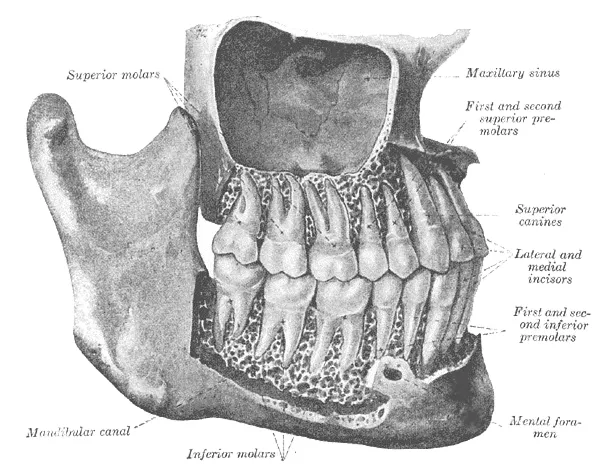
Gray's Anatomy plate of the permanent teeth with bone removed, labeling the incisors, canines, premolars and molars in both jaws
Plate II · LeadPermanent teeth in the jaws with the roots exposedHenry Vandyke Carter · Public domain

In this plate, we cover

  1. 01 Tooth numbering systems 6 facts
  2. 02 Eruption of the primary teeth 9 facts
  3. 03 Eruption of the permanent teeth 12 facts
  4. 04 Incisors 6 facts
  5. 05 Canines 4 facts
  6. 06 Premolars 7 facts
  7. 07 Molars 9 facts
  8. 08 Primary dentition 7 facts
  9. 09 Occlusion 7 facts
  10. 10 Developmental anomalies 9 facts

01 · 6 facts

Tooth numbering systems

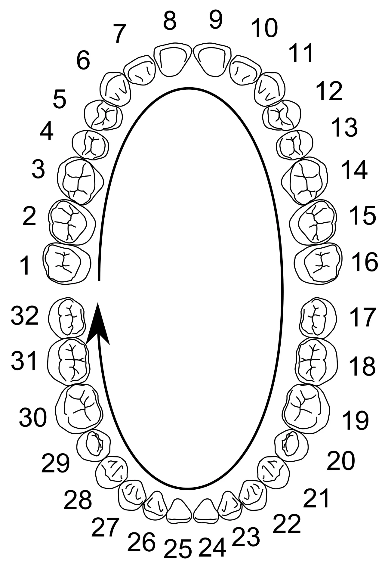
Three systems name teeth. The Universal system numbers permanent teeth 1-32, starting at the maxillary right third molar, and letters primary teeth A through T. The FDI system uses two digits, quadrant first and tooth second, so Universal #8 is 11 and #19 is 36. Palmer notation places 1-8 or A-E inside a quadrant bracket.

  1. Universal: permanent teeth 1-32 start at the maxillary right third molar, go across to 16, drop to 17 (mandibular left third molar) and end at 32.S1
  2. Universal primary teeth: A-J maxillary right to left, K-T mandibular left to right.S1
  3. FDI two-digit system: first digit is the quadrant (1-4 permanent, 5-8 primary), second is the tooth (1-8). Tooth 11 = #8; 36 = #19.T6
  4. Palmer notation uses a quadrant bracket with 1-8 for permanent and A-E for primary teeth.T6
All 6 facts on tooth numbering systems
Chart of the permanent teeth numbered 1 to 32 using the Universal Numbering System
Fig. II.1Universal Numbering SystemKaligula · CC BY-SA 3.0

02 · 9 facts

Eruption of the primary teeth

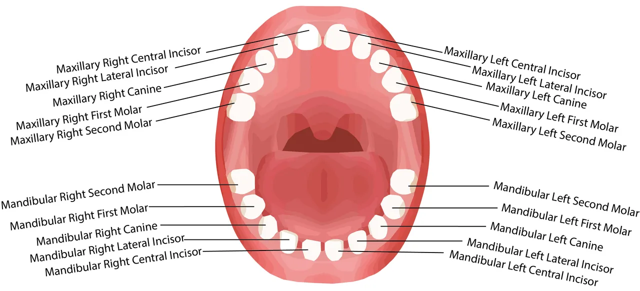
The 20 primary teeth begin erupting at about 6 months, usually led by the mandibular central incisors. The order is central incisor, lateral incisor, first molar, canine, then second molar, which arrives last at roughly 23-33 months. The primary dentition period lasts until about age 6, when the mixed dentition begins and runs to about 12.

  1. Primary teeth: 20 in all, beginning to erupt at about 6 months; mandibular central incisors usually come first.S6
  2. Mandibular primary central incisor erupts 6-10 months; maxillary central 8-12 months.S6
  3. Primary lateral incisors erupt 9-13 months (maxillary) and 10-16 months (mandibular).S6
  4. Primary first molars erupt 13-19 months (maxillary) and 14-18 months (mandibular).S6
All 9 facts on eruption of the primary teeth
Diagram of an open mouth with each primary tooth labeled by name
Fig. II.2Names of the primary teethGregory david baker · CC BY-SA 4.0

03 · 12 facts

Eruption of the permanent teeth

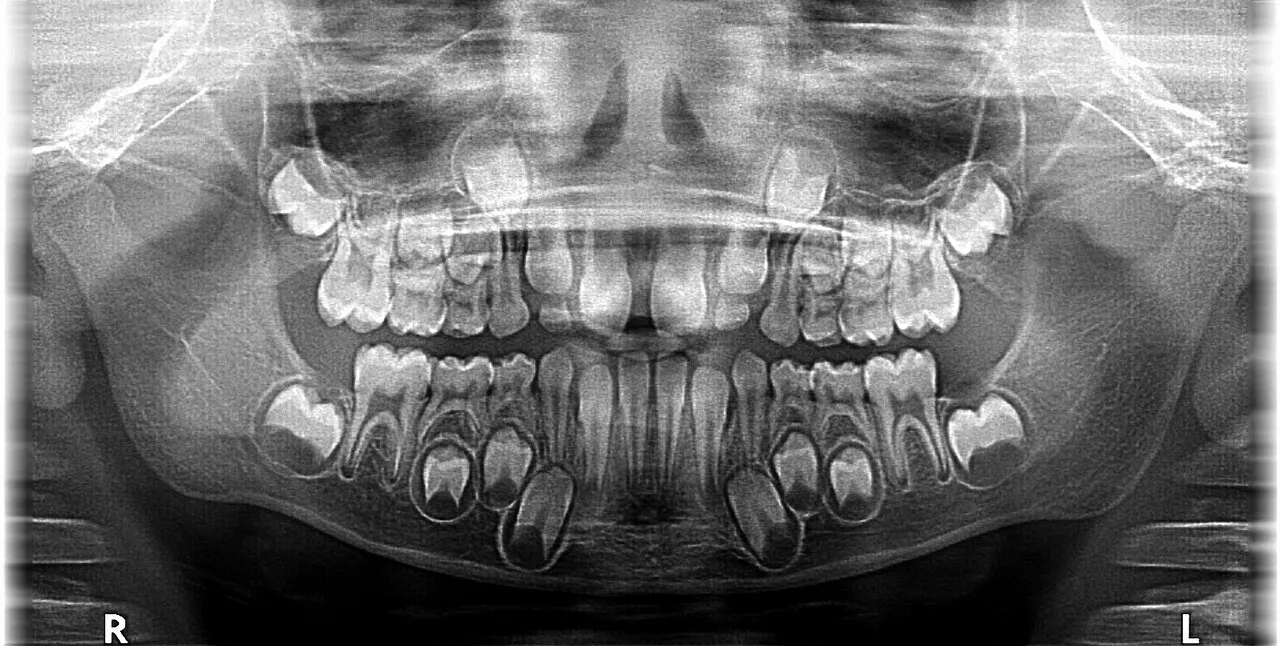
Permanent teeth arrive from about age 6, when the first molars and mandibular central incisors erupt, through the third molars at 17-21. Mandibular teeth usually lead their maxillary partners, and the maxillary canine (11-12 years) typically follows the premolars. Molars have no primary predecessor, while incisors, canines and premolars replace primary teeth.

  1. First permanent molars and mandibular central incisors erupt at 6-7 years.S6
  2. Maxillary central incisors 7-8 years; maxillary lateral incisors 8-9 years; mandibular lateral incisors 7-8 years.S6
  3. Mandibular canine 9-10 years; maxillary canine 11-12 years.S6
  4. Maxillary first premolar 10-11 years; maxillary second premolar 10-12 years.S6
All 12 facts on eruption of the permanent teeth
Panoramic radiograph of a child showing primary teeth with the developing permanent teeth beneath them
Fig. II.3Panoramic image of a mixed dentitionGang65 · CC BY-SA 4.0

04 · 6 facts

Incisors

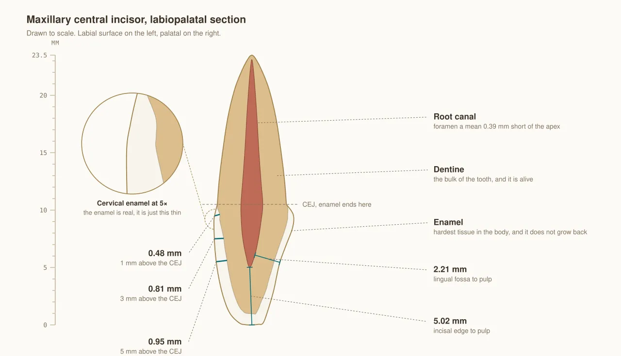
Incisors are the front cutting teeth. The maxillary central has the widest crown of any anterior tooth, the maxillary lateral is the most variable and may be peg-shaped or missing, and the mandibular central is the smallest permanent tooth. Mandibular incisor roots are flattened side to side with proximal concavities, and newly erupted incisors show three mamelons.

  1. Maxillary central incisor: widest crown mesiodistally of any anterior tooth; mesial incisal angle is sharp, distal is rounded.T6
  2. Mamelons (3) appear on newly erupted incisors and wear away with function.T6
  3. Maxillary lateral incisor: most variable anterior tooth; may be peg-shaped or congenitally missing; often has a lingual pit.T6
  4. Mandibular central incisor: smallest permanent tooth and the most bilaterally symmetrical.T6
All 6 facts on incisors
Scale drawing of a maxillary central incisor cut labiolingually, showing enamel, dentin and pulp
Fig. II.4Maxillary central incisor in sectionKhalid Aletaibi · CC BY 4.0

05 · 4 facts

Canines

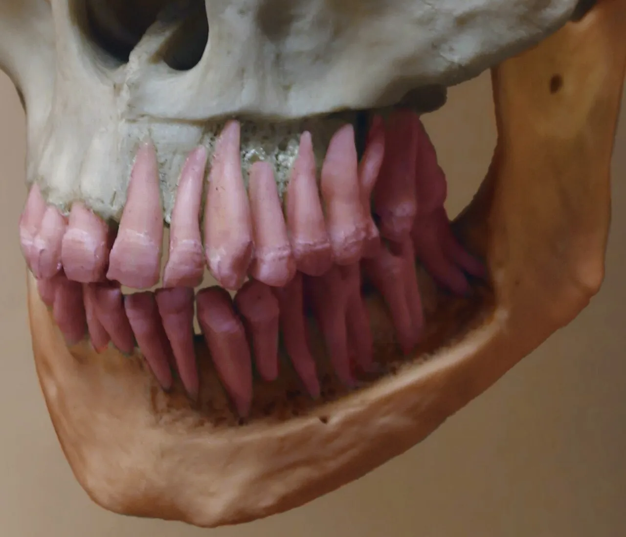
Canines sit at the corners of the arch and guide the jaw in side-to-side movement. The maxillary canine has the longest root in the mouth, about 17 mm, which raises the canine eminence over it. On both canines the mesial cusp ridge is shorter than the distal, and the mandibular canine is the anterior tooth most likely to have a split root.

  1. The maxillary canine has the longest root in the mouth, about 17 mm on average.T6
  2. Canines form the "corner" of the arch and the canine eminence; they guide lateral excursions (canine guidance).T6
  3. On the maxillary canine the mesial cusp ridge is shorter than the distal; the same is true on the mandibular canine.T6
  4. The mandibular canine is the anterior tooth most likely to have a bifurcated root (labial and lingual).T6
All 4 facts on canines
Skull with the alveolar bone removed so the full length of each root is visible, including the long canine roots
Fig. II.5Alveolar bone cut away to show the rootsGrey Geezer · CC BY-SA 4.0

06 · 7 facts

Premolars

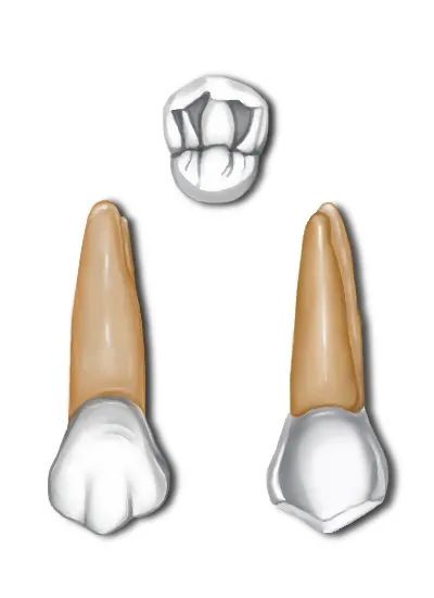
The eight premolars replace the primary molars and have no counterpart in the primary dentition. The maxillary first premolar usually has two roots and a prominent mesial concavity on crown and root; the maxillary second has one root and nearly equal cusps. The mandibular first premolar's small lingual cusp makes it look like a canine, and the mandibular second most often has three cusps.

  1. Premolars are 8 in all, replace the primary molars and are absent from the primary dentition.T6
  2. Maxillary first premolar: two roots (buccal and palatal) in most cases; a prominent mesial crown and root concavity (mesial developmental depression).T6
  3. Maxillary first premolar: buccal cusp is longer than the lingual; the mesial cusp ridge of the buccal cusp is longer than the distal (unlike the canine).T6
  4. Maxillary second premolar: usually one root; cusps nearly equal in height.T6
All 7 facts on premolars
Drawings of a maxillary first premolar from two sides with an occlusal view above showing its buccal and lingual cusps
Fig. II.6Maxillary first premolarΑνώνυμος Βικιπαιδιστής · CC BY 3.0

07 · 9 facts

Molars

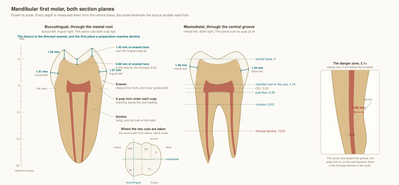
Maxillary first molars have four main cusps, often a cusp of Carabelli, an oblique ridge and three roots, with the palatal root longest and furcations opening buccally, mesially and distally. Mandibular first molars have five cusps and two widely separated roots, mesial and distal, and their short root trunk means the buccal and lingual furcations are exposed early as attachment is lost.

  1. Maxillary first molar: 4 major cusps plus the cusp of Carabelli on the mesiolingual cusp; mesiolingual is the largest cusp.T6
  2. Maxillary first molar: three roots; palatal is longest; furcations open buccally, mesially and distally.T6
  3. The oblique ridge (mesiolingual to distobuccal cusp) is found only on maxillary molars.T6
  4. Maxillary second molar: smaller, often lacks Carabelli; distolingual cusp is smaller or absent.T6
All 9 facts on molars
Scale drawings of a mandibular first molar cut in two planes showing enamel, dentin and pulp
Fig. II.7Mandibular first molar cross sectionsKhalid Aletaibi · CC BY 4.0

08 · 7 facts

Primary dentition

Primary teeth have whiter, thinner enamel (about 1 mm), a pronounced cervical ridge and large pulps with high pulp horns. Primary molar roots are thin and flare wide to make room for the premolars forming beneath them. Each primary second molar resembles the permanent first molar of the same arch, and primate spaces next to the canines are normal.

  1. Primary teeth have whiter, thinner enamel (about 1 mm), a pronounced cervical ridge, and larger pulps with high horns.S43
  2. Primary molar roots are thin and flare widely to make room for the premolar crowns.S43
  3. The primary maxillary second molar resembles the permanent maxillary first molar.S43
  4. The primary mandibular second molar resembles the permanent mandibular first molar (3 nearly equal buccal cusps).S43
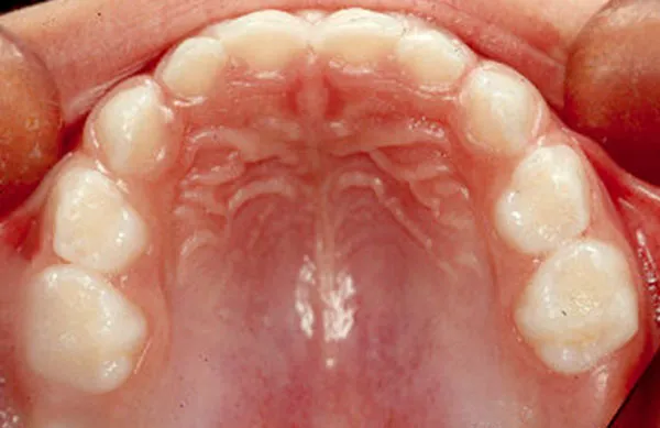
All 7 facts on primary dentition
Occlusal view of a child's upper arch of primary teeth
Fig. II.8Maxillary primary archwww.ortodoncia.ws · CC BY-SA 3.0

09 · 7 facts

Occlusion

Occlusion is how the arches meet. In Angle Class I the mesiobuccal cusp of the maxillary first molar sits in the buccal groove of the mandibular first molar; in Class II the mandibular molar is distal to that spot, and in Class III it is mesial. Normal overjet and overbite are each about 2-3 mm, and the curves of Spee and Wilson shape the occlusal plane.

  1. Angle Class I: the MB cusp of the maxillary first molar sits in the buccal groove of the mandibular first molar.T6
  2. Angle Class II: the mandibular molar is distal to that position (retrognathic profile). Division 1 has flared maxillary incisors; Division 2 has retroclined centrals.T6
  3. Angle Class III: the mandibular molar is mesial (prognathic profile).T6
  4. Normal overjet (horizontal) and overbite (vertical) are about 2-3 mm, or about 20-30% overlap for overbite.T6
All 7 facts on occlusion
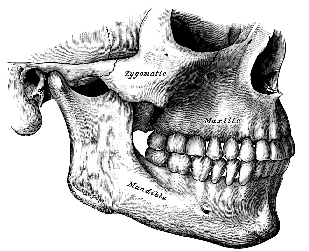
Gray's Anatomy plate of the skull from the side showing the maxillary and mandibular teeth meeting in occlusion
Fig. II.9Teeth in occlusion, side viewHenry Vandyke Carter · Public domain

10 · 9 facts

Developmental anomalies

Developmental anomalies change tooth number, shape or structure. Third molars are the teeth most often missing, and a mesiodens between the maxillary centrals is the most common extra tooth. Gemination keeps the normal tooth count while fusion lowers it, concrescence joins roots by cementum, and a talon cusp adds an extra cusp on the lingual of an anterior tooth.

  1. Hypodontia most often affects third molars, then mandibular second premolars and maxillary lateral incisors.T5
  2. A mesiodens is a supernumerary tooth between the maxillary central incisors; it is the most common supernumerary.T5
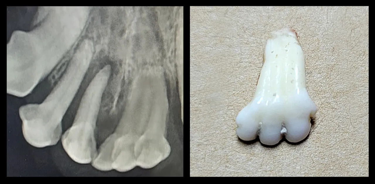
  3. Gemination: one tooth germ tries to split (normal tooth count). Fusion: two germs join (one fewer tooth).T5
  4. Concrescence is the joining of two teeth by cementum only, after root formation.T5
All 9 facts on developmental anomalies
Radiograph (left) and extracted specimen (right) of two tooth crowns joined into one tooth
Fig. II.10Fused teeth on a radiograph and after extractionMarialeegRVT · CC BY-SA 4.0

Watch

Film II.1How to Number Teeth CONFIDENTLY! Palmer, FDI, and Universal Tooth Numbering Systems Made SimpleDental Clarafication with Dr. Clara · YouTube · plays from youtube-nocookie.com
Film II.2Tooth Numbering Systems / Dental Notations (Universal, Palmers and FDI tooth numbering systems)DentalManiaK · YouTube · plays from youtube-nocookie.com