Atlas · Plate III · Scientific Basis
Histology and Embryology
Histology and embryology explain what the tissues you scale, probe and polish are made of and how they formed. The area covers the basic tissue types, the stages of tooth development, the structure of enamel, dentin, pulp and cementum, the periodontal ligament and alveolar bone, the gingiva and oral mucosa, and how the face and palate come together before birth.
On the NBDHE it is tested in the Scientific Basis section, and it sits underneath many clinical answers: why enamel cannot repair itself, why exposed dentin is sensitive, why the junctional epithelium is where periodontal inflammation starts, and why some cysts arise from leftover epithelium. It also explains how cleft lip and cleft palate happen.
Blueprint Scientific Basis for Dental Hygiene Practice · Anatomic Sciences: histology and embryology · 50 facts

In this plate, we cover
01 · 5 facts
Cells and tissues
The body is built from four basic tissues: epithelial, connective, muscle and nervous. Keratinized oral epithelium has basal, prickle, granular and keratin layers, while parakeratinized epithelium, common on attached gingiva, keeps nuclei at the surface. Desmosomes join cells to each other, hemidesmosomes join them to a basal lamina, and fibroblasts are the main cells of the PDL and gingival connective tissue.
- Four basic tissues: epithelial, connective, muscle, nervous.S49
- Keratinized epithelium layers from deep to surface: basal, prickle (spinous), granular, keratin (cornified).S52
- Parakeratinized epithelium keeps nuclei in the surface layer; it is common on attached gingiva.S52
- Hemidesmosomes attach cells to a basal lamina; desmosomes attach cells to each other.S52
02 · 7 facts
Tooth development
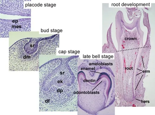
Teeth begin forming around the sixth to seventh week in utero and pass through initiation, bud, cap, bell, apposition and maturation. The enamel organ makes enamel, the dental papilla becomes dentin and pulp, and the dental follicle becomes cementum, PDL and bone. Hertwig's epithelial root sheath shapes the root, and its leftover rests of Malassez can later form cysts.
- Teeth arise from ectoderm (enamel) and neural crest-derived ectomesenchyme (dentin, pulp, cementum, PDL).S52
- Primary tooth development begins around the 6th-7th week in utero with the dental lamina.S52
- Stages: initiation, bud, cap, bell, apposition, maturation.S52
- Enamel organ, dental papilla and dental sac (follicle) form at the cap stage; papilla becomes dentin and pulp, follicle becomes cementum, PDL and alveolar bone.S52
03 · 7 facts
Enamel
Enamel is about 96% mineral, mostly hydroxyapatite, and is the hardest tissue in the body. It is laid down as rods that run from the dentinoenamel junction to the surface. Because ameloblasts are lost once the tooth erupts, enamel cannot regrow. Growth lines called lines of Retzius end on the surface as perikymata, and enamel is thickest over the cusps.
- Enamel is about 96% inorganic hydroxyapatite by weight; it is the hardest tissue in the body.S44
- Ameloblasts form enamel; they are lost after eruption, so enamel cannot regenerate.S52
- The enamel rod (prism) is the basic structural unit; it runs from the DEJ to the surface.S52
- Lines of Retzius are incremental growth lines; they end on the surface as perikymata.S52

04 · 7 facts
Dentin and pulp
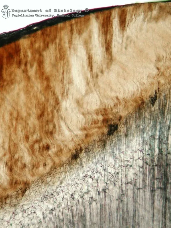
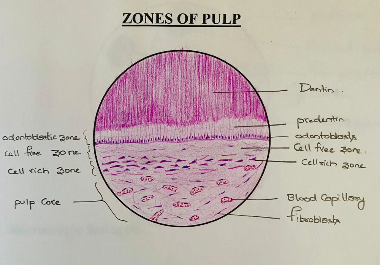
Dentin is about 70% mineral and is made by odontoblasts, which line the pulp for life and send processes into the dentinal tubules. It keeps forming slowly as secondary dentin and faster as tertiary dentin after injury. Fluid moving in open tubules explains dentin hypersensitivity. The pulp has four zones, from the odontoblast layer inward to the pulp core.
- Dentin is about 70% inorganic, 20% organic (mostly type I collagen) and 10% water by weight.S44
- Odontoblasts line the pulp and send processes into dentinal tubules; they persist for life.S52
- Primary dentin forms before root completion; secondary dentin forms slowly for life; tertiary (reparative/reactionary) dentin forms in response to injury.S52
- Sclerotic dentin has tubules filled with mineral, which reduces sensitivity.S52

05 · 9 facts
Cementum, PDL and alveolar bone
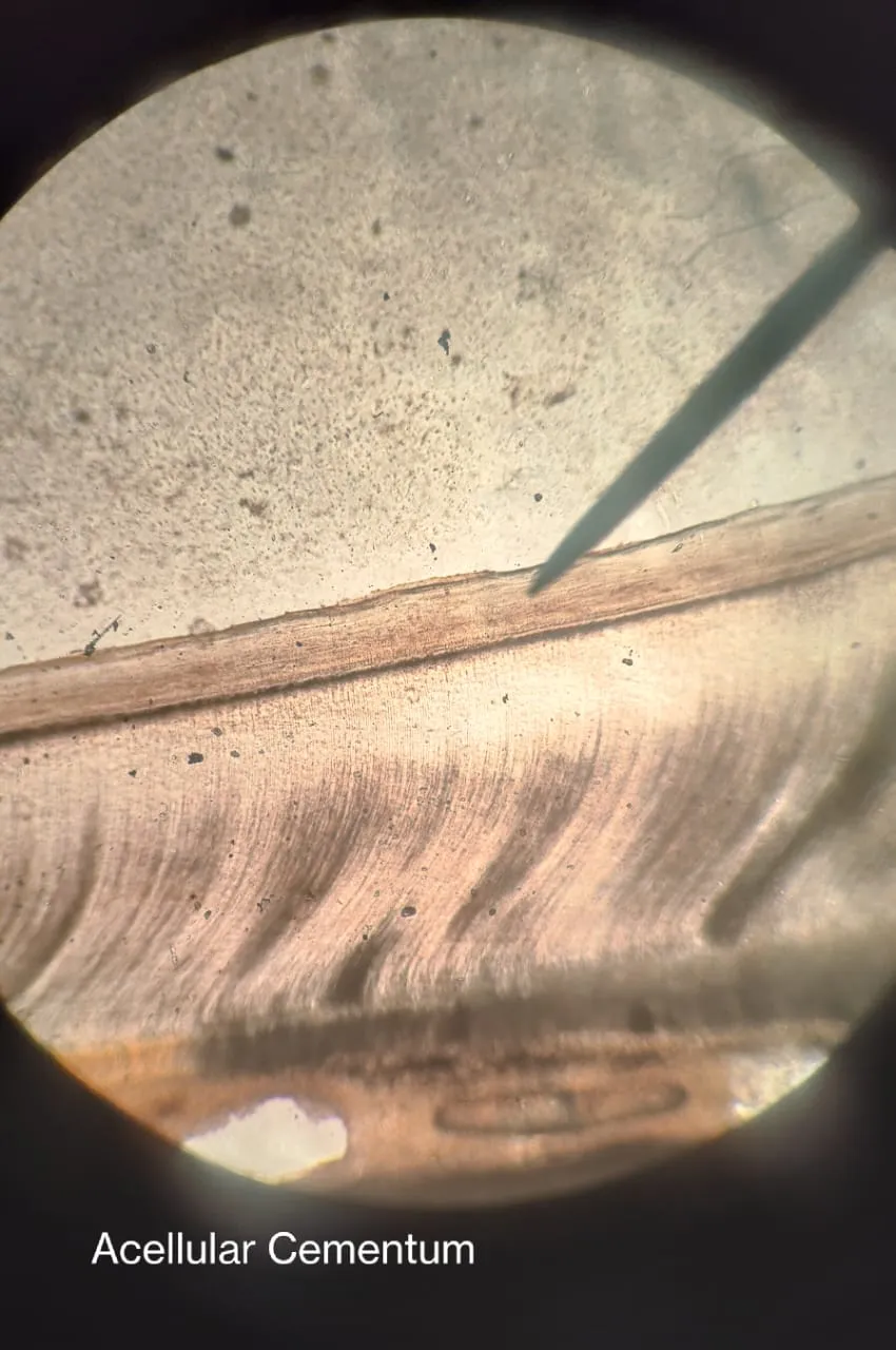
Cementum is the softest of the tooth's hard tissues, about 45-50% mineral, with no blood or nerve supply; acellular cementum covers the cervical root and cellular cementum the apical third. PDL collagen fibers, anchored as Sharpey fibers, hold the tooth in bone, and the oblique group, the largest, resists biting force. The lamina dura is the socket lining seen on radiographs.
- Cementum is about 45-50% inorganic; it is avascular and has no innervation.S40
- Acellular (primary) cementum covers the cervical root; cellular (secondary) cementum is on the apical third and furcations.S40
- Sharpey fibers are PDL collagen fibers embedded in cementum and alveolar bone.S40
- The PDL is about 0.15-0.38 mm wide and thinnest at the middle third of the root.S40

06 · 9 facts
Gingiva and oral mucosa
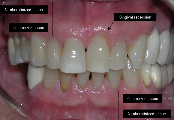
Oral mucosa is masticatory (keratinized gingiva and hard palate), lining (nonkeratinized cheeks, floor of mouth and soft palate) or specialized (the dorsum of the tongue). Healthy gingiva is coral pink, firm and knife-edged, and the mucogingival junction marks where attached gingiva ends. The junctional epithelium seals to the tooth, renews in about 4-6 days and is where periodontal inflammation begins.
- Oral mucosa types: masticatory (gingiva, hard palate; keratinized), lining (buccal, labial, floor of mouth, ventral tongue, soft palate; nonkeratinized), specialized (dorsal tongue).S52
- Attached gingiva is keratinized and bound to bone; it is widest in the maxillary incisor area and narrowest at the mandibular premolars/canines (facial).S41
- The mucogingival junction separates attached gingiva from alveolar mucosa.S41
- Free gingival groove separates free from attached gingiva (present in about one-third of adults).S41

07 · 6 facts
Face and palate development
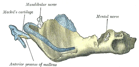
The face forms between the fourth and eighth weeks from five swellings: the frontonasal process and the paired maxillary and mandibular processes. The primary palate comes from the fused medial nasal processes, and the secondary palate from palatal shelves that fuse around weeks 7-12. When these fusions fail, the result is cleft lip or cleft palate. The first pharyngeal arch builds the jaws and chewing muscles.
- The face develops from 5 primordia: frontonasal process, paired maxillary processes, paired mandibular processes, between the 4th and 8th weeks.S52
- The primary palate (premaxilla) comes from the fused medial nasal processes; the secondary palate comes from the palatal shelves of the maxillary processes.S52
- Palatal shelves fuse between about weeks 7 and 12 in utero.S52
- Cleft lip is more common in males and unilateral on the left; isolated cleft palate is more common in females.T5

Watch