Atlas · Plate I · Scientific Basis
Head and Neck Anatomy
Head and neck anatomy is the map behind most of what a hygienist does: the extraoral and intraoral exam, palpating lymph nodes and muscles, and placing local anesthetic near the right nerve without entering a vessel. It covers the skull and mandible, the temporomandibular joint, the muscles of mastication and facial expression, the cranial nerves, the blood supply, lymph nodes, salivary glands and the fascial spaces that infection can travel through.
On the NBDHE it sits in the Scientific Basis section under anatomic sciences, and it returns in clinical cases. Expect questions that tie a structure to a consequence: which nodes drain a given tooth, why a PSA needle placed too deep can cause a hematoma, why anesthetic deposited in the parotid causes temporary facial paralysis, and how a mandibular molar infection reaches the neck.
Blueprint Scientific Basis for Dental Hygiene Practice · Anatomic Sciences: head and neck anatomy · 66 facts

In this plate, we cover
- 01 Skull and facial bones 9 facts
- 02 Temporomandibular joint 7 facts
- 03 Muscles of mastication and expression 10 facts
- 04 Cranial nerves 11 facts
- 05 Blood supply and venous drainage 6 facts
- 06 Lymph nodes 7 facts
- 07 Salivary glands 6 facts
- 08 Fascial spaces and spread of infection 4 facts
- 09 Tongue and oral landmarks 6 facts
01 · 9 facts
Skull and facial bones
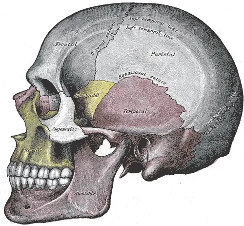
The skull has 22 bones, 8 cranial and 14 facial, and the mandible is the only one that moves. For hygienists the payoff is the foramina: the mandibular foramen on the inner ramus, the mental foramen near the apex of the mandibular second premolar, and the infraorbital, greater palatine and incisive foramina used as injection landmarks.
- The skull has 22 bones: 8 cranial and 14 facial.S49
- The mandible is the only movable skull bone; the maxillae are paired and fuse at the intermaxillary suture.S49T4
- The mandibular foramen sits on the medial ramus, about midway between the anterior and posterior borders; the lingula overlaps it.S15
- The mental foramen usually lies below the apex of the mandibular second premolar (or between the premolars).S68
02 · 7 facts
Temporomandibular joint
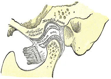
The TMJ joins the mandibular condyle to the articular fossa and eminence of the temporal bone. Its disc splits the joint into a lower space that hinges and an upper space that glides, so opening starts with rotation and then the condyle slides forward. Normal opening is about 40-50 mm, and a click on opening usually means the disc sits too far forward.
- The TMJ is a ginglymoarthrodial joint: it both hinges (rotates) and glides (translates).T4
- The articular disc divides the joint into an upper (gliding/translation) and a lower (hinge/rotation) space.T4
- The joint is formed by the mandibular condyle and the articular fossa and eminence of the temporal bone.T4
- The lateral pterygoid inserts partly on the disc and the condylar neck and pulls the condyle forward.T4

03 · 10 facts
Muscles of mastication and expression
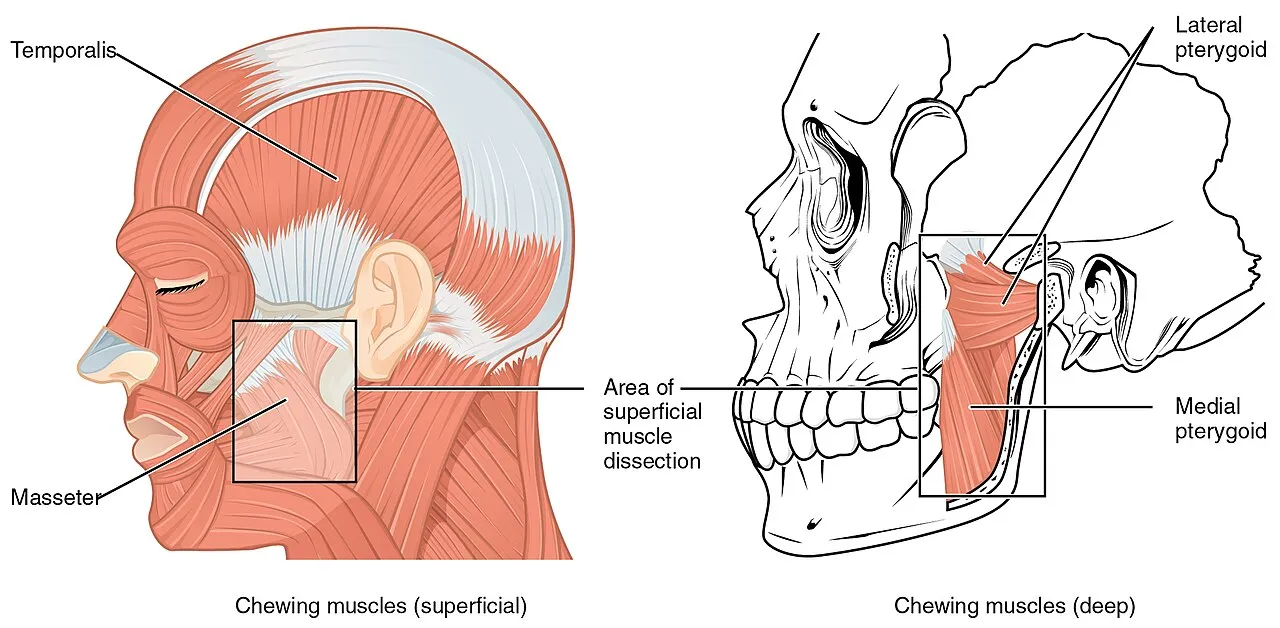
Four muscles of mastication, all supplied by V3, move the mandible. The masseter, temporalis and medial pterygoid close the jaw, while the lateral pterygoid protrudes it and helps open. The muscles of facial expression, including the buccinator and orbicularis oris, are all supplied by the facial nerve (CN VII). Palpate the masseter on both sides during the extraoral exam.
- Muscles of mastication: masseter, temporalis, medial pterygoid, lateral pterygoid; all are supplied by V3.S45
- Elevators (closers): masseter, temporalis, medial pterygoid.T4
- The inferior head of the lateral pterygoid is the main protruder and helps open; one side alone moves the jaw to the opposite side.T4
- The posterior fibers of the temporalis retract the mandible.T4

04 · 11 facts
Cranial nerves
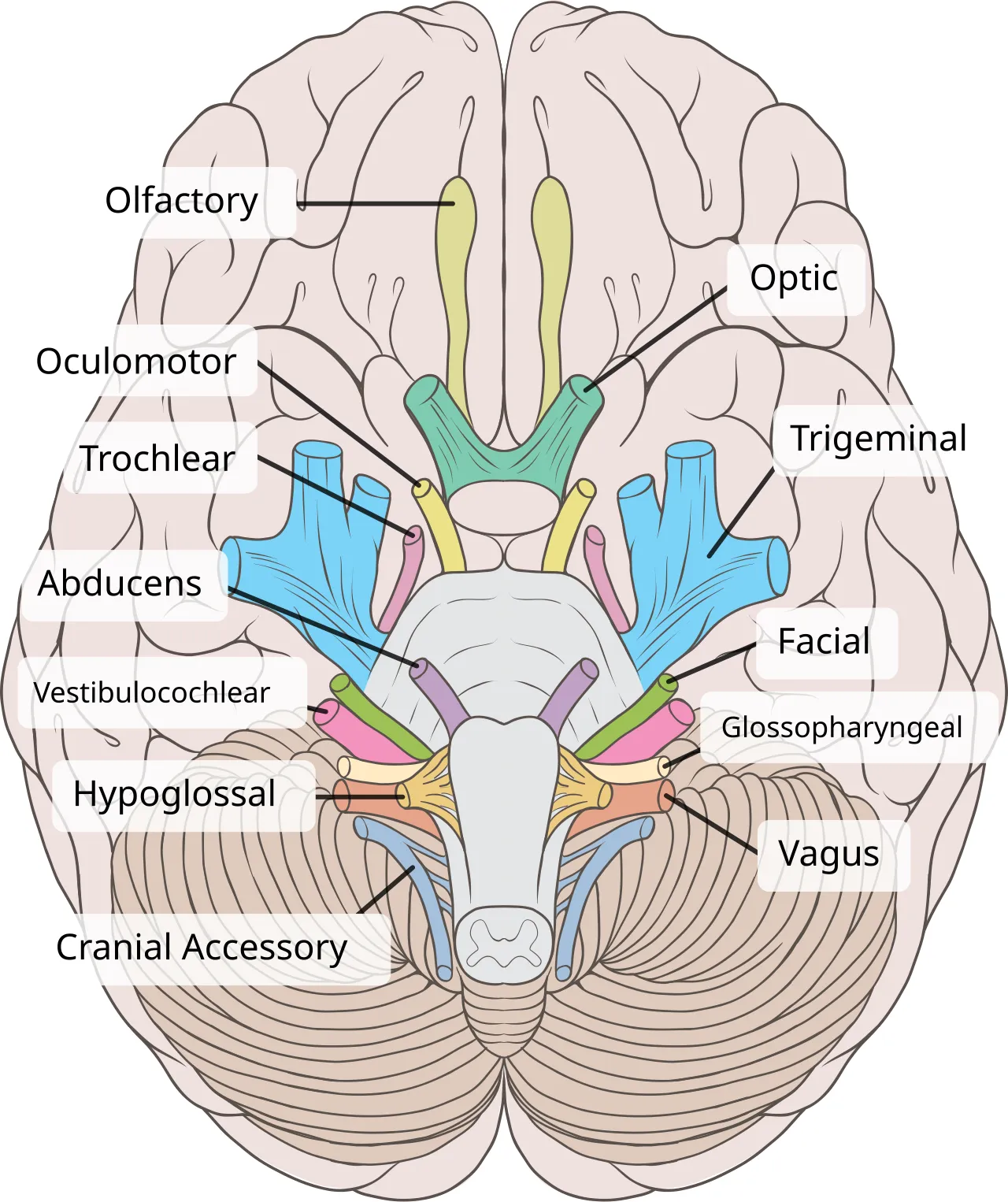
Twelve pairs of cranial nerves leave the brain, and the trigeminal (V) is the main sensory nerve of the face and teeth: V2 serves the maxillary teeth, V3 the mandibular teeth plus the chewing muscles. The facial nerve (VII) runs through the parotid to the muscles of expression, CN IX carries taste from the back third of the tongue, and CN XII moves the tongue.
- There are 12 pairs of cranial nerves; the trigeminal (V) is the largest and is the main sensory nerve of the face and teeth.S49
- V1 ophthalmic is sensory only; V2 maxillary is sensory only; V3 mandibular is both sensory and motor.S45
- V2 exits the skull through the foramen rotundum; V3 exits through the foramen ovale.S45
- V3 motor fibers supply mastication muscles plus mylohyoid, anterior digastric, tensor tympani and tensor veli palatini.S45

05 · 6 facts
Blood supply and venous drainage
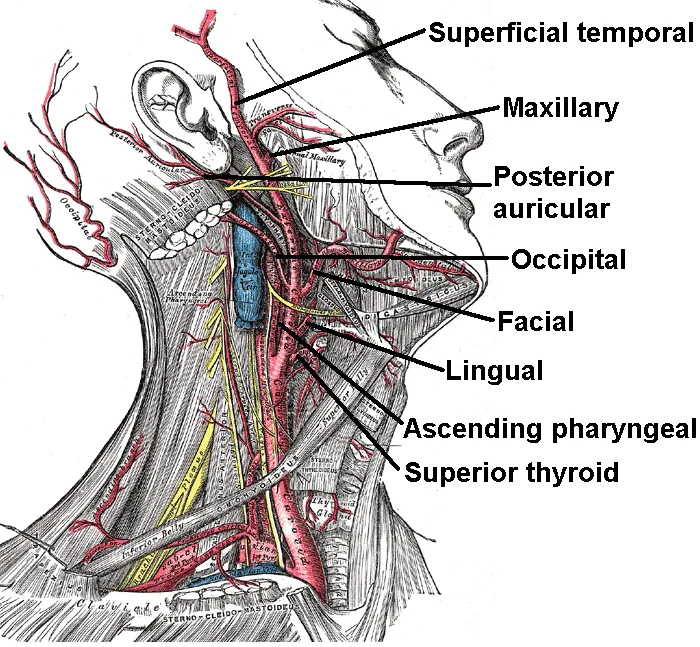
The external carotid artery feeds the face and mouth, and its maxillary branch gives off the alveolar arteries that supply the teeth. The veins matter for safety: the pterygoid plexus around the lateral pterygoid is where a too-deep PSA needle causes a hematoma, and valveless facial veins connect to the cavernous sinus, so infection above the upper lip can spread inside the skull.
- The external carotid artery supplies the face and mouth; the maxillary artery is one of its two terminal branches.T4
- The maxillary artery gives the inferior alveolar, posterior superior alveolar, infraorbital, greater palatine and other branches.T4
- The lingual artery supplies the tongue; the facial artery is palpable at the anterior border of the masseter on the lower mandible.T4
- The pterygoid plexus of veins lies around the lateral pterygoid; a PSA needle inserted too deep can cause a hematoma there.S16

06 · 7 facts
Lymph nodes
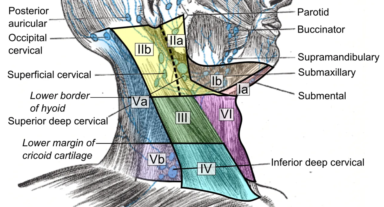
Lymph nodes filter drainage from the teeth and face, so they are palpated in every extraoral exam, comparing sides and working from superior to inferior. Submental nodes drain the mandibular incisors and tongue tip, submandibular nodes drain most other teeth, and all of them empty into the deep cervical chain. Tender, soft, mobile nodes suggest infection; hard, fixed, painless nodes are a cancer red flag.
- Superficial nodes include occipital, retroauricular, anterior auricular, superficial parotid, facial, submental and submandibular nodes.T4
- Submental nodes drain the mandibular incisors, chin, center of lower lip, floor of mouth and tongue tip.T4
- Submandibular nodes drain most maxillary and mandibular teeth (except mandibular incisors), the cheeks, upper lip and the anterior tongue.T4
- Maxillary third molars can drain to the superior deep cervical nodes.T4

07 · 6 facts
Salivary glands
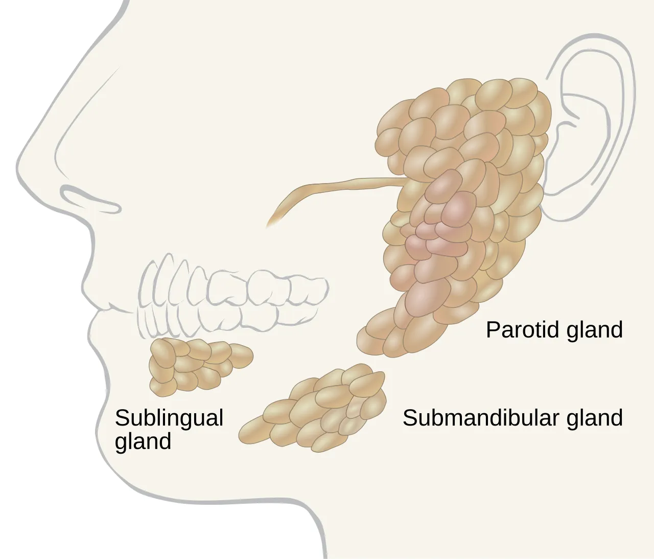
Three pairs of major glands make saliva: the parotid (largest and purely serous), the submandibular (mixed) and the sublingual (smallest and mostly mucous). The parotid duct opens on the cheek opposite the maxillary second molar, and the submandibular duct opens at the sublingual caruncle; that gland forms most salivary stones. Minor glands are scattered through the oral mucosa.
- Three paired major glands: parotid (largest, purely serous), submandibular (mixed, mostly serous), sublingual (smallest, mixed, mostly mucous).S49
- The parotid duct (Stensen) opens at the parotid papilla on the buccal mucosa opposite the maxillary second molar.T4
- The submandibular duct (Wharton) opens at the sublingual caruncle beside the lingual frenum.T4
- About 80-90% of salivary stones form in the submandibular gland, because its duct runs uphill and its saliva is thicker.T5

08 · 4 facts
Fascial spaces and spread of infection
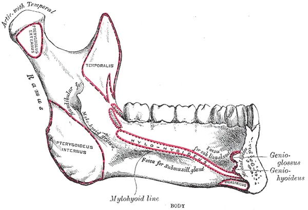
Fascial spaces are connected planes between tissues, so a dental infection can track from a tooth into the neck. Mandibular second and third molar infections tend to reach the submandibular space because their apices sit below the mylohyoid line. Ludwig angina, a cellulitis of the submandibular, sublingual and submental spaces on both sides, can close off the airway.
- Fascial spaces link up, so dental infections can spread from the teeth into the neck.T4
- Ludwig angina is bilateral cellulitis of the submandibular, sublingual and submental spaces; it can block the airway.T4
- Mandibular second and third molar infections tend to spread into the submandibular space (apices below the mylohyoid line).T4
- Maxillary canine infections can spread through the canine space to the cavernous sinus.T4

09 · 6 facts
Tongue and oral landmarks
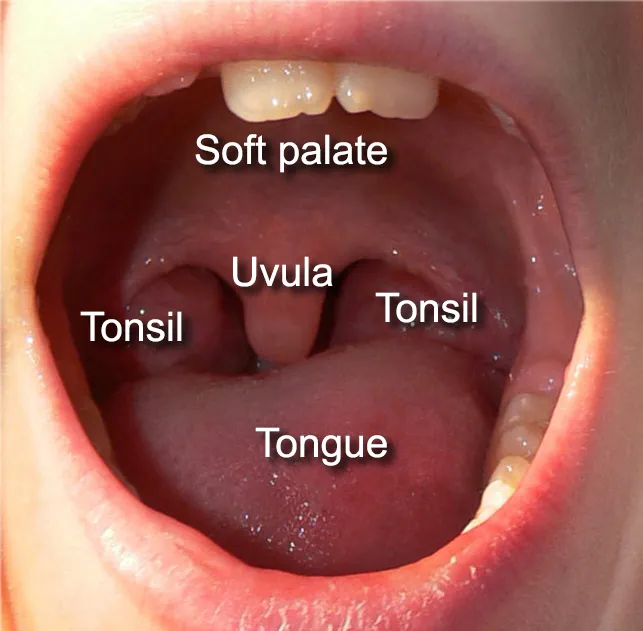
The dorsum of the tongue carries four kinds of papillae: numerous filiform papillae with no taste buds, red-dot fungiform papillae with taste buds, foliate papillae on the back edges, and 8-12 circumvallate papillae in a V in front of the sulcus terminalis. Knowing normal variants such as Fordyce granules and the linea alba keeps you from charting them as lesions.
- The circumvallate papillae (8-12) form a V in front of the sulcus terminalis.S52
- Filiform papillae are the most numerous and have no taste buds; foliate papillae are on the posterolateral tongue.S52
- Fungiform papillae look like red dots on the dorsum and contain taste buds.S52
- The foramen cecum marks where the thyroid gland began to descend.S52

Watch