Atlas · Plate X · Clinical Services
Periodontology
Periodontology is the study of the tissues that hold teeth in place, the gingiva, periodontal ligament, cementum and alveolar bone, and of the biofilm-driven diseases that destroy them. It sits at the center of hygiene practice: you probe, chart, classify and debride, then decide when to reevaluate and when to refer.
Periodontal disease management is a core NBDHE clinical subarea and runs through the patient cases, which pair charts with radiographs. Know the 2017 AAP/EFP classification well enough to assign stage and grade from CAL, bone loss, tooth loss, smoking and HbA1c. Also expect NSPT outcomes, local and host-modulating agents, and the line between peri-implant mucositis and peri-implantitis.
Blueprint Provision of Clinical Dental Hygiene Services · Periodontal disease management · 64 facts

In this plate, we cover
01 · 6 facts
The periodontium
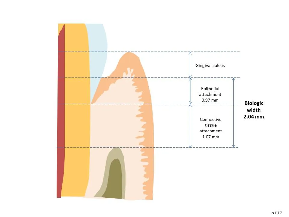
The periodontium is four tissues: gingiva, periodontal ligament, cementum and alveolar bone. A healthy sulcus probes about 0.5-3 mm, and crevicular fluid increases with inflammation. The lamina dura lines each socket. Root concavities, such as the mesial of the maxillary first premolar, trap deposits and make instrumentation harder.
- The periodontium = gingiva, PDL, cementum and alveolar bone.S40
- Healthy sulcus depth is about 0.5-3 mm (histologic depth is about 0.69 mm).S40T2
- Gingival crevicular fluid increases with inflammation and carries neutrophils, antibodies and enzymes.S40
- Alveolar bone proper (lamina dura) lines the socket; supporting bone includes cortical plates and cancellous (trabecular) bone.S40

02 · 8 facts
Etiology and pathogenesis
Plaque biofilm starts gingivitis and periodontitis, but most tissue breakdown comes from the host's own response, through enzymes and mediators such as MMPs, IL-1, TNF-alpha and PGE2. Smoking is the strongest modifiable risk factor and diabetes roughly triples risk. Local factors like calculus, overhangs and open contacts help biofilm stay in place.
- Plaque biofilm is the primary cause of gingivitis and periodontitis; the host response causes most of the tissue destruction.S38
- Page and Schroeder lesions: initial (2-4 days), early (4-7 days), established (2-3 weeks, plasma cells), advanced (periodontitis, bone loss).S38
- Destructive mediators include MMPs (collagenases), IL-1, TNF-alpha and PGE2.S38
- Smoking is the strongest modifiable risk factor; smokers have about 2-7 times the periodontitis risk and less bleeding (vasoconstriction masks inflammation).S38

03 · 19 facts
2017 classification: staging and grading
The 2017 AAP/EFP system replaced the old chronic and aggressive labels with a single periodontitis described by stage, extent and grade. Stage I-IV reflects severity and complexity from CAL, bone loss and tooth loss. Grade A-C reflects the rate of progression, using bone loss divided by age, smoking and HbA1c. Gingivitis means 10% or more bleeding sites without attachment loss.
- Periodontal health: under 10% bleeding sites and probing depths 3 mm or less on an intact periodontium.S12S13
- Dental plaque-induced gingivitis: 10% or more bleeding sites with probing depths 3 mm or less and no attachment loss.S12
- Localized gingivitis is 10-30% bleeding sites; generalized is over 30%.S12
- Periodontitis case: interdental CAL at 2 or more nonadjacent teeth, or buccal/oral CAL of 3 mm or more with pocketing over 3 mm at 2 or more teeth.S12S11

04 · 7 facts
Nonsurgical periodontal therapy
Nonsurgical therapy removes biofilm and calculus from root surfaces, coaches self-care and controls risk factors. Expect less bleeding and moderate pockets that shrink by about 1-2 mm, healing mainly by a long junctional epithelium. Reevaluate about 4-6 weeks later and refer residual bleeding pockets of 5 mm or more, furcations and progressing cases.
- Nonsurgical periodontal therapy (NSPT) = biofilm and calculus removal (scaling and root debridement), self-care coaching, risk-factor control, plus adjuncts as needed.S38
- Modern root debridement removes calculus and biofilm without deliberately removing cementum.T3
- Expected NSPT results: bleeding falls; moderate pockets (4-6 mm) drop about 1-2 mm; deep pockets (7 mm or more) drop about 2-3 mm through shrinkage and gain in clinical attachment.S38T3
- Healing after NSPT is mostly a long junctional epithelium, not new attachment.T3
05 · 8 facts
Chemotherapeutic agents
Chlorhexidine 0.12%, essential oil and cetylpyridinium chloride rinses help control gingivitis; chlorhexidine stains and is inactivated by sodium lauryl sulfate. Locally delivered minocycline, doxycycline gel and the chlorhexidine chip go into pockets of 5 mm or more after scaling. Subantimicrobial-dose doxycycline modulates the host by inhibiting collagenase rather than killing bacteria.
- Chlorhexidine gluconate 0.12%: 15 mL rinse for 30 s twice daily; substantivity about 8-12 hours; stains teeth, alters taste, increases supragingival calculus.T2
- Wait about 30 min between toothpaste with sodium lauryl sulfate and chlorhexidine because SLS inactivates it.T2
- Essential oil rinses and cetylpyridinium chloride rinses are ADA-accepted antigingivitis agents.T2
- Minocycline microspheres (1 mg per site) are placed into pockets of 5 mm or more after scaling.T3
06 · 6 facts
Surgical therapy
When pockets persist after nonsurgical care, flap and osseous surgery give access and recontour bone, guided tissue regeneration uses a membrane to regrow support, and gingival or connective tissue grafts treat thin tissue and recession. The hygienist supports aftercare: periodontal dressings protect the wound for about a week, and sutures usually come out in 7-10 days.
- Gingivectomy removes excess gingiva (e.g., drug-induced enlargement) when there is adequate attached gingiva.T3
- Flap surgery gives access to roots and bone; osseous surgery reshapes bone.T3
- Guided tissue regeneration uses a barrier membrane to keep epithelium out so PDL and bone can regrow.T3
- Free gingival graft (palate donor) increases keratinized tissue; connective tissue graft covers recession roots better.T3

07 · 6 facts
Implants and peri-implant disease
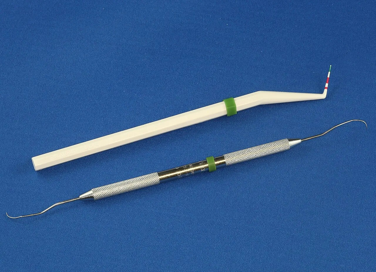
Implants have no periodontal ligament, so peri-implant tissues seal less well and have less blood supply. Probe with light force and treat bleeding or suppuration as a warning sign. Peri-implant mucositis is reversible inflammation; peri-implantitis adds progressive bone loss. Use implant-safe instruments and glycine or erythritol powders, and avoid APF fluoride.
- Implants have no PDL; peri-implant tissue has parallel collagen fibers, a weaker seal and less blood supply than teeth.S12
- Implant probing with light force is recommended; plastic probes are optional. Bleeding or suppuration on probing signals disease.S12
- Use plastic, resin, titanium or implant-safe tips; avoid steel instruments that scratch titanium and APF fluoride that etches it.T3T2
- Glycine or erythritol air polishing is effective and safe for implant biofilm.T3

08 · 4 facts
Occlusal trauma
Primary occlusal trauma is excessive force on a healthy periodontium; secondary trauma is normal or excessive force on a periodontium already reduced by disease. Look for mobility, fremitus, a widened PDL space, wear facets and pain on biting. Trauma does not start periodontitis on its own but can speed bone loss where inflammation is present.
- Primary occlusal trauma: excessive forces on a normal periodontium. Secondary: normal or excess forces on a reduced periodontium.S12
- Signs: mobility, fremitus, widened PDL space, wear facets, pain on biting.T3
- Fremitus is vibration felt when the patient taps the teeth together, usually on maxillary teeth.T3
- Occlusal trauma does not cause periodontitis by itself but can speed bone loss where inflammation exists.S12
Watch