Atlas · Plate XII · Clinical Services
Patient Assessment and Charting
Patient assessment is the information-gathering stage of the dental hygiene process: the health history and vital signs, the extraoral and intraoral exam, hard-tissue charting, periodontal charting and the indices that put numbers on plaque, bleeding and disease. Everything planned afterward, from appointment length to referrals, rests on getting these findings right.
Assessing patient characteristics is a core NBDHE clinical subarea, and every patient case opens with a history box, vital signs, charts and images to interpret. Practice calculating clinical attachment level, naming G.V. Black classes, grading furcations and mobility, matching each index to what it measures, and writing a dental hygiene diagnosis that states both the problem and its cause.
Blueprint Provision of Clinical Dental Hygiene Services · Assessing patient characteristics · 43 facts

In this plate, we cover
01 · 6 facts
Medical history and vital signs
Review and update the health history, medications and allergies at every visit. Anticoagulants, bisphosphonates and immunosuppressants may call for a medical consult or a change in care, but only the prescriber stops a drug. Elective care is usually deferred when blood pressure reaches about 180/110, and patients on drugs that cause orthostatic hypotension should sit up slowly.
- Review and update the medical history at every visit, including medications, allergies and recent changes.T2
- Patients on bisphosphonates, anticoagulants or immunosuppressants need consults or modifications.S57S67
- Routine dental cleaning can proceed for warfarin patients when the INR is in range (usually under about 3.5 for minor procedures); check a recent INR.S57
- Do not tell patients to stop antithrombotic drugs; the prescriber decides.S57
02 · 7 facts
Extraoral and intraoral exam
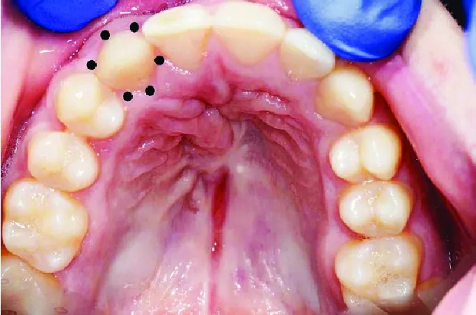
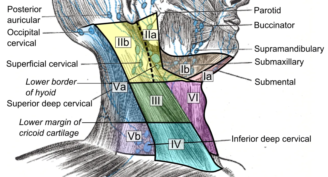
The extraoral exam checks skin, eyes, TMJ, muscles of mastication, lymph nodes, thyroid, salivary glands and lips; the intraoral exam covers the mucosa, tongue, floor of the mouth, palate, oropharynx and gingiva. Match the palpation type to the site, pull the tongue out with gauze to see its lateral borders, and describe lesions by size, color, shape, surface and base.
- Extraoral exam: skin, eyes, TMJ, muscles of mastication, lymph nodes, thyroid, salivary glands, lips.T2
- The thyroid moves up when the patient swallows; palpate it from behind or in front during swallowing.T2
- Intraoral exam: labial and buccal mucosa, vestibules, tongue (dorsal, lateral, ventral), floor of mouth, hard and soft palate, oropharynx, gingiva.T2
- Grasp the tongue with gauze to examine the lateral borders, the highest-risk site for oral cancer.T2

03 · 8 facts
Periodontal charting
Probing depth runs from the gingival margin to the pocket base, while clinical attachment level runs from the CEJ to the base and shows how much support is lost. Add recession to the probing depth when the margin sits apical to the CEJ, and subtract when it sits coronal. Charts also record mobility (Miller) and furcation involvement (Glickman or Hamp).
- Probing depth: gingival margin to the base of the pocket. Clinical attachment level: CEJ to the base of the pocket.T3
- CAL = PD + recession when the margin is apical to the CEJ; CAL = PD when the margin is at the CEJ; CAL = PD minus the distance from the CEJ to the margin when the margin is coronal to the CEJ.T3
- Example: PD 5 mm with 2 mm recession = CAL 7 mm. PD 6 mm with the margin 2 mm above the CEJ = CAL 4 mm.T3
- Width of attached gingiva = width of keratinized gingiva (margin to MGJ) minus probing depth.T3
04 · 6 facts
Hard-tissue charting
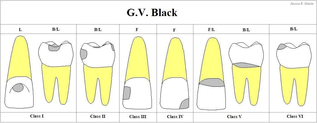
Hard-tissue charting records caries, restorations and wear. G.V. Black classes locate lesions, from Class I pits and fissures to Class VI cusp tips and incisal edges. Wear takes four forms: attrition, abrasion, erosion and abfraction. ICDAS grades caries from 0 (sound) to 6 (extensive cavity), and root caries clusters in older adults with recession and dry mouth.
- G.V. Black Class I: pits and fissures (occlusal surfaces of posterior teeth, buccal/lingual pits, lingual pits of maxillary incisors).T2
- Class II: proximal surfaces of premolars and molars. Class III: proximal surfaces of incisors and canines without the incisal angle. Class IV: proximal of anteriors including the incisal angle.T2
- Class V: gingival third of facial or lingual surfaces. Class VI: incisal edges or cusp tips.T2
- Attrition is tooth-to-tooth wear; abrasion is wear by a foreign object (e.g., toothbrush); erosion is chemical; abfraction is a stress-related notch at the cervical area.T2

05 · 11 facts
Indices
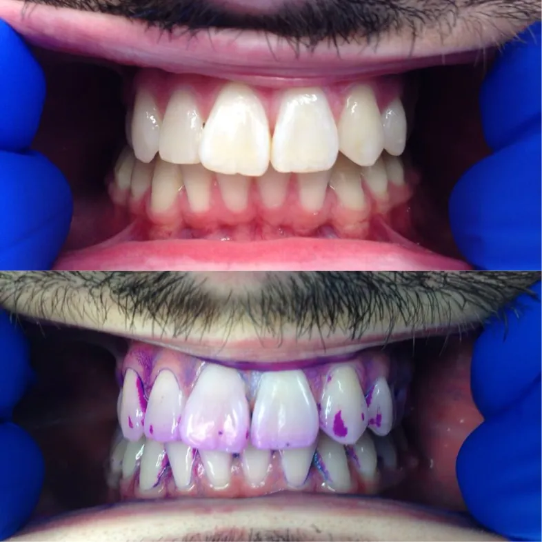
Indices turn findings into scores you can track. The O'Leary Plaque Control Record aims for 10% or less, OHI-S combines debris and calculus scores on six index surfaces, the Gingival Index grades inflammation from 0 to 3, and DMFT counts lifetime caries experience. Plaque and gingival indices are reversible, while DMF and attachment measures are irreversible.
- Plaque Control Record (O'Leary): percentage of surfaces with plaque, 4 surfaces per tooth; a goal of 10% or less.T2
- Patient Hygiene Performance (Podshadley and Haley): same 6 index teeth as OHI-S, each surface divided into 5 areas, score 0-5 per tooth.T2
- OHI-S index teeth: facial of #3, #8, #14, #24 and lingual of #19 and #30.T2
- Debris and calculus indices each score 0-3; OHI-S totals 0-6; 0-1.2 good, 1.3-3.0 fair, 3.1-6.0 poor.T2

06 · 5 facts
Care plan and DH diagnosis
The process runs assessment, diagnosis, planning, implementation, evaluation and documentation. A dental hygiene diagnosis names a problem the hygienist can treat plus its cause. Goals should be measurable and time-bound, and case presentation covers findings, options, risks, costs and the outcome of no treatment before consent. A typical NSPT plan ends with reevaluation at 4-6 weeks.
- The dental hygiene process: assessment, dental hygiene diagnosis, planning, implementation, evaluation, documentation (ADPIE + D).T2
- A dental hygiene diagnosis names a problem the hygienist is educated and licensed to treat, plus its cause (e.g., gingival bleeding related to inadequate biofilm removal).T2
- Goals in the care plan should be measurable and time-bound (e.g., bleeding under 10% in 6 weeks).T2
- Case presentation explains findings, options, risks, costs, and the outcome of no treatment, then gets informed consent.T2

Watch