Atlas · Plate VII · Scientific Basis
General and Oral Pathology
General and oral pathology covers how tissue reacts to injury, how cells adapt or become neoplastic, and how to recognize the lesions found during an oral cancer screening: white, red and pigmented lesions, ulcers and blisters, soft-tissue lumps, jaw cysts and tumors, and the oral signs of inherited and systemic disease.
Chairside, the working rule is simple: a suspicious lesion that has not resolved about two weeks after removing a possible cause is referred for biopsy. The NBDHE tests lesion recognition in clinical photos and radiographs, the far higher risk of erythroplakia compared with leukoplakia, the usual sites of oral squamous cell carcinoma, and classic jaw lesions such as the periapical cyst, the dentigerous cyst and the odontogenic keratocyst.
Blueprint Scientific Basis for Dental Hygiene Practice · Pathology (general and oral) · 63 facts
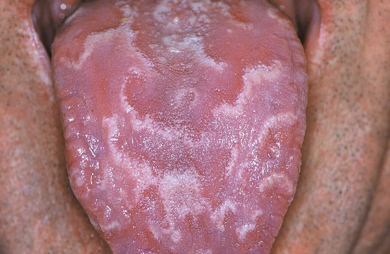

In this plate, we cover
01 · 6 facts
Inflammation and repair
Inflammation shows five cardinal signs: redness, heat, swelling, pain and loss of function. Histamine from mast cells opens vessels early, neutrophils dominate acute inflammation, and lymphocytes, plasma cells and macrophages take over when it becomes chronic. Clean wound edges heal by primary intention, while a gap fills with granulation tissue by secondary intention. An abscess is a localized collection of pus.
- Five cardinal signs of inflammation: redness (rubor), heat (calor), swelling (tumor), pain (dolor), loss of function.S49
- Acute inflammation is dominated by neutrophils; chronic inflammation by lymphocytes, plasma cells and macrophages.T5
- Histamine from mast cells causes early vasodilation and increased permeability.T5
- Healing by primary intention closes clean wound edges; secondary intention fills a gap with granulation tissue.T5
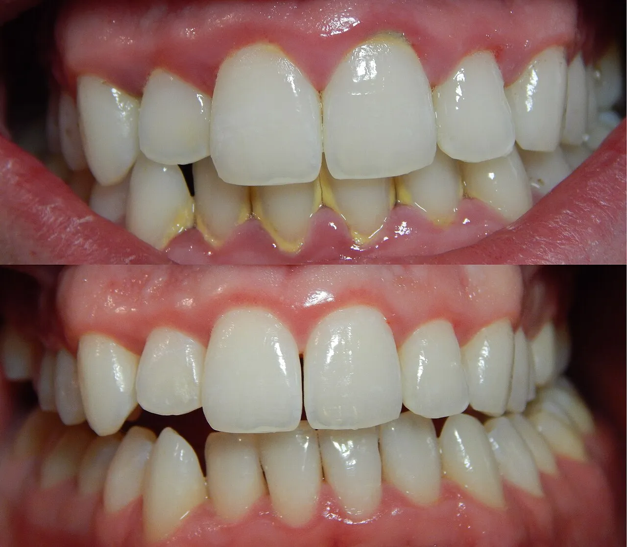

02 · 6 facts
Cell adaptation and neoplasia
Cells adapt by multiplying (hyperplasia), enlarging (hypertrophy), shrinking (atrophy) or changing into another mature cell type (metaplasia). Dysplasia is disordered growth with atypical cells; when it spans the full epithelium without breaking the basement membrane, it is carcinoma in situ. Benign tumors grow slowly and stay put, malignant tumors invade and spread, and TNM staging records tumor size, nodes and metastasis.
- Hyperplasia is more cells; hypertrophy is bigger cells; atrophy is smaller cells or tissue.T5
- Metaplasia replaces one mature cell type with another; dysplasia is disordered growth with atypical cells and can precede cancer.T5
- Carcinoma in situ is full-thickness dysplasia that has not broken through the basement membrane.T5
- Benign tumors are encapsulated, slow and do not metastasize; malignant tumors invade and spread.T5
03 · 7 facts
White lesions
Many white changes are harmless: linea alba, frictional keratosis and leukoedema, which fades when the cheek is stretched. Others deserve a closer look: smokeless tobacco keratosis where the tobacco is held, nicotine stomatitis on a pipe smoker's palate, the lacy Wickham striae of lichen planus on both cheeks, and EBV-related hairy leukoplakia on the lateral tongue. Pseudomembranous candidiasis wipes off; leukoplakia cannot be rubbed off.
- Frictional keratosis, linea alba and leukoedema are benign white changes; leukoedema disappears when the mucosa is stretched.T5
- Smokeless tobacco keratosis forms where the tobacco is held (usually the mandibular vestibule).T5
- Nicotine stomatitis: white palate with red dots (inflamed minor salivary gland ducts), linked to pipe smoking and hot drinks.T5
- Oral lichen planus is a T cell-mediated condition; the reticular form (Wickham striae) is the most common; erosive forms are painful.S46
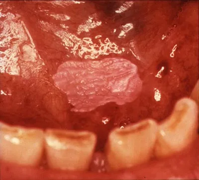

04 · 6 facts
Red and pigmented lesions
Red lesions range from the reactive pyogenic granuloma, which bleeds easily and is called a pregnancy tumor when it appears in pregnancy, to the gingiva-only peripheral giant cell granuloma and the hemangioma, which blanches under pressure. Erythroplakia is the red patch with the highest cancer risk. Among pigmented lesions, the amalgam tattoo is most common, physiologic pigmentation is symmetric, and oral melanoma is rare.
- Pyogenic granuloma is a reactive red lump that bleeds easily, often on the gingiva; it is called a "pregnancy tumor" in pregnancy.T5
- Peripheral giant cell granuloma occurs only on the gingiva or alveolar ridge.T5
- Hemangioma blanches with pressure (diascopy).T5
- Amalgam tattoo is the most common oral pigmented lesion; it is flat, blue-gray and does not change.T5
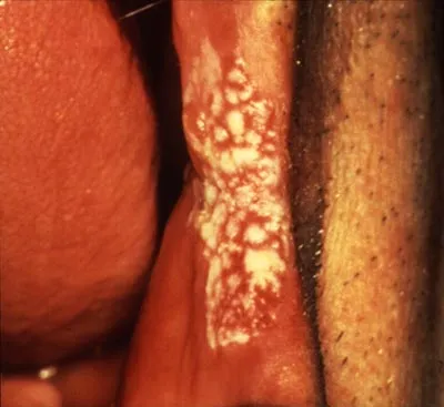

05 · 6 facts
Ulcers and vesicles
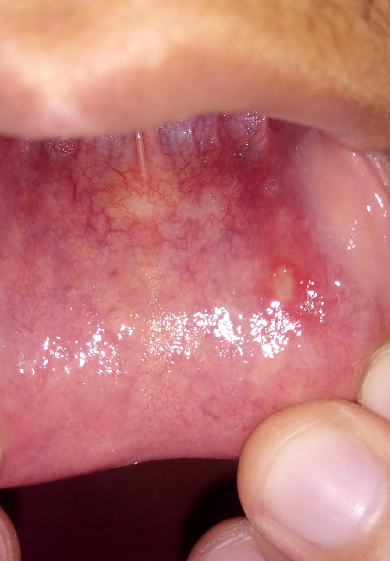
Minor aphthous ulcers are under 1 cm, form on movable nonkeratinized mucosa and heal in 7-14 days without scarring; major aphthae are larger, last weeks and scar. Recurrent herpes favors keratinized tissue such as the palate, attached gingiva and lip. Primary herpetic gingivostomatitis brings fever and painful gingivitis with vesicles, and erythema multiforme causes target skin lesions and crusted lips.
- Minor aphthous ulcers are under 1 cm and heal in 7-14 days without scarring; major aphthae are over 1 cm, last weeks and scar.T5
- Aphthous ulcers occur on nonkeratinized (movable) mucosa; recurrent intraoral herpes occurs on keratinized mucosa (palate, attached gingiva).T5
- Primary herpetic gingivostomatitis has fever, malaise and painful generalized gingivitis with vesicles.T5
- Traumatic ulcers resolve within about 2 weeks once the cause is removed.T5

06 · 6 facts
Soft-tissue masses
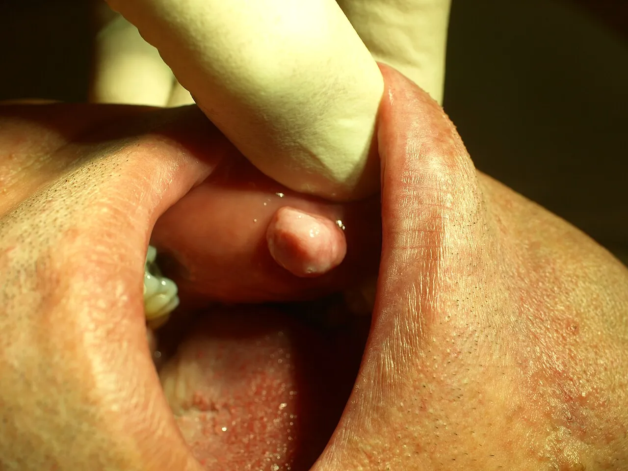
The irritation fibroma, a firm nodule along the bite line of the cheek, is the most common oral soft-tissue mass. A mucocele is a mucus-filled swelling, usually on the lower lip, from a ruptured minor salivary gland duct; in the floor of the mouth it is called a ranula. Papillomas are cauliflower-like, epulis fissuratum follows a denture flange, and tori are benign bony growths.
- Irritation fibroma is the most common oral soft tissue mass; it usually forms on the buccal mucosa along the bite line.T5
- Mucocele: most often on the lower lip from a ruptured minor salivary gland duct.T5
- Ranula is a large mucocele in the floor of the mouth from the sublingual gland.T5
- Papilloma: cauliflower-like, pedunculated, often on the soft palate or tongue (HPV 6, 11).T5

07 · 9 facts
Cysts and odontogenic tumors
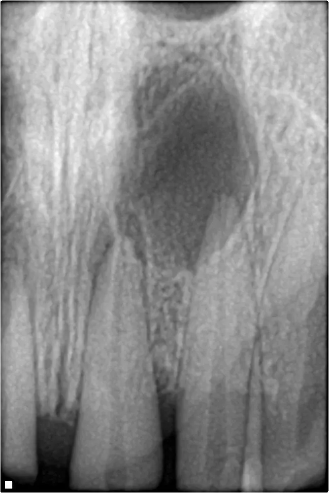
The periapical cyst at the apex of a nonvital tooth is the most common jaw cyst, and the dentigerous cyst surrounds the crown of an unerupted tooth, usually a mandibular third molar. The odontogenic keratocyst recurs often and, when multiple, suggests Gorlin syndrome. The odontoma is the most common odontogenic tumor, and the ameloblastoma forms a soap-bubble radiolucency in the posterior mandible.
- Periapical (radicular) cyst: most common jaw cyst; arises from rests of Malassez at the apex of a nonvital tooth.S47
- Dentigerous cyst: second most common odontogenic cyst; attaches at the CEJ of an unerupted tooth, usually a mandibular third molar.S47
- Odontogenic keratocyst: high recurrence (often cited around 25-30%); multiple OKCs suggest Gorlin syndrome.S47
- Lateral periodontal cyst: between roots of mandibular canine/premolar area, on a vital tooth.S47

08 · 10 facts
Oral cancer
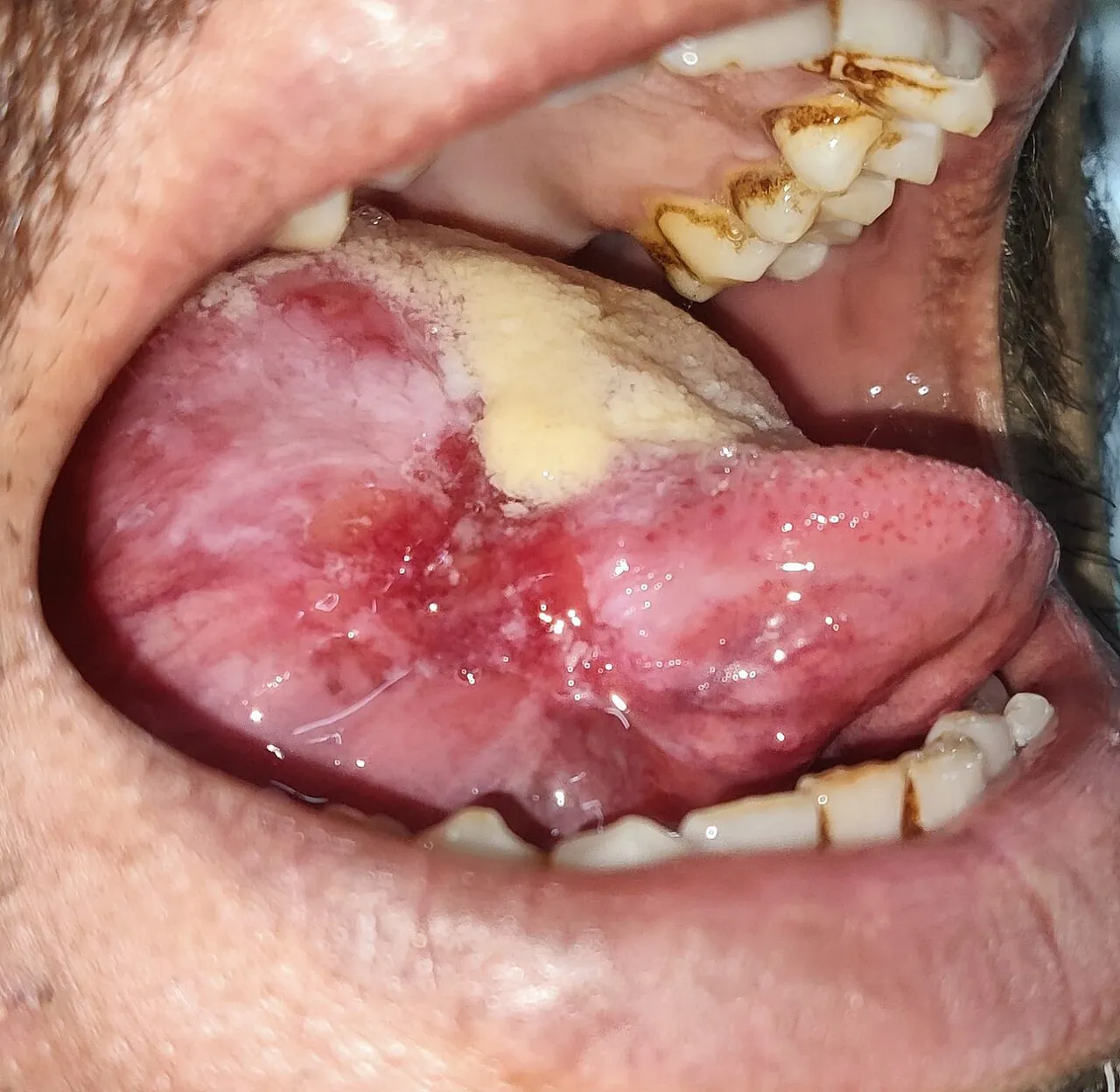
Squamous cell carcinoma makes up about 90% of oral cancers. Tobacco and alcohol together raise risk sharply, HPV-16 drives oropharyngeal cancer and sun exposure drives lower lip cancer. The posterolateral and ventral tongue and the floor of the mouth are the usual intraoral sites. Survival drops when cancer is found late, so a suspicious lesion lasting about two weeks after removing any cause goes to biopsy.
- Squamous cell carcinoma makes up about 90% of oral cancers.S58
- Main risk factors: tobacco and alcohol (synergistic), HPV-16 for oropharynx, sun exposure for lip cancer.S58
- Most common intraoral sites: posterolateral border and ventral tongue, then floor of the mouth.S58T5
- Lower lip SCC is linked to sun exposure; actinic cheilitis is its precursor.T5

09 · 7 facts
Genetic and systemic conditions
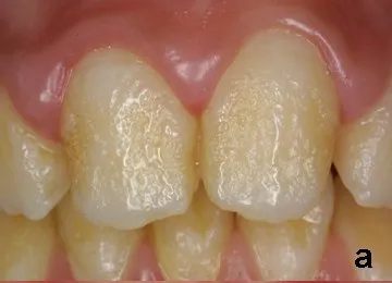
Some conditions show up first in the mouth. Down syndrome brings early severe periodontitis, macroglossia and a fissured tongue; ectodermal dysplasia gives missing or cone-shaped teeth; amelogenesis imperfecta affects only enamel, while dentinogenesis imperfecta causes opalescent teeth with obliterated pulps. Addison disease adds oral pigmentation, Peutz-Jeghers syndrome perioral freckles, and Sjögren syndrome a dry mouth and dry eyes.
- Down syndrome (trisomy 21): early severe periodontitis, macroglossia, fissured tongue, delayed eruption, hypodontia, heart defects.T5
- Ectodermal dysplasia: missing or cone-shaped teeth, sparse hair, few sweat glands.T5
- Amelogenesis imperfecta affects enamel only; dentinogenesis imperfecta gives opalescent teeth and obliterated pulps.T5
- Cleidocranial dysplasia: missing or underdeveloped clavicles, many unerupted supernumerary teeth.T5

Watch